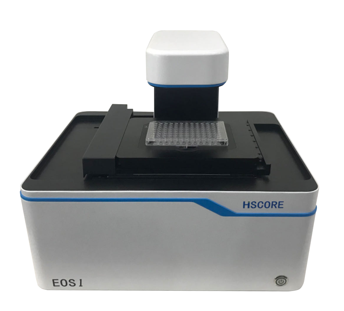
Eos1 Living Cell Imaging and Analysis System
High Throughput Cell Imaging Analysis System

 location:Home-Product--Living Cell Imaging Analysis System-Eos1 Living Cell Imaging and Analysis System
High Throughput Cell Imaging Analysis System
Performance characteristics:
Optical system:
Olympus objective lens, optimized optical system, provides high quality cell images
Objective configuration:
Automatic objective turntable for 4X, 10X, 20X, 40X objectives
Imaging mode:
bright field, phase contrast, monochromatic fluorescence, multicolor fluorescence (red, green and blue), Z-Stacking, full-hole imaging
Software algorithms:
AI algorithms based on powerful artificial intelligence provide more accurate cell division and analysis
Long-term observation:
The instrument can be placed in the CO2 incubator to achieve long-term time lapse imaging observation
Application field:
Organoids:
Culture observation of organoids, monochromatic and multicolor fluorescence analysis of organoids
Cell health and vitality analysis:
Cell count, cell proliferation, cell cycle, apoptosis,3D tumor spheres
Cell morphology and movement:
cell migration, scratch test, chemotaxis
Cell function:
immune cell killing, cell phagocytosis, angiogenesis

LED excitation: 365nm, 500nm,575nm,645nm(option)
Emission: 455nm,520nm,595nm, 705nm(option)
Transmitted modes: Bright field , Phase contrast
Objective turret: 4 position
Objectives: 4X,10X,20X,40X(option)
Automatic XY stage: 115 X 86mm, 1um repeatabliity
Motorized Z: 14mm,100nm step,Z-stack
Camera: high sensitivity monocherome 5MP comos
please contact us by mail.